

A behind-the-scenes peek into the S&T laboratory that is critical to our nation’s defense against biological threats and our COVID-19 pandemic response.
-
Image
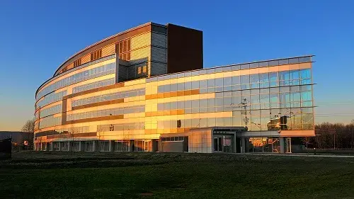
NBACC, located at Fort Detrick in Frederick, MD, was founded after 9/11 to defend the nation against biological threats, manmade and natural.
-
Image
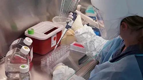
NBACC scientist working in Biosafety Level 3 containment in a Class II biological safety cabinet performs dispensing of samples using a micropipette, which measures very small volumes of liquid.
-
Image
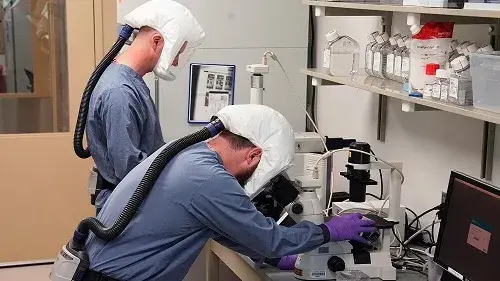
NBACC scientists working in Biosafety Level 3 containment examine cells with a microscope to detect infection at a cellular level.
-
Image
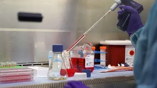
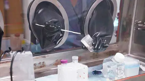
NBACC scientist working in a Class II biosafety cabinet within Biosafety Level 3 containment uses a serological pipette to transfer fluids for research studies
-
Image
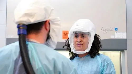
An NBACC safety officer confers with an NBACC scientist prior to an experiment being conducted in a Biosafety Level 3 lab. Powered air purifying respirators are used to protect the workers from potential aerosol exposures.
-
Image
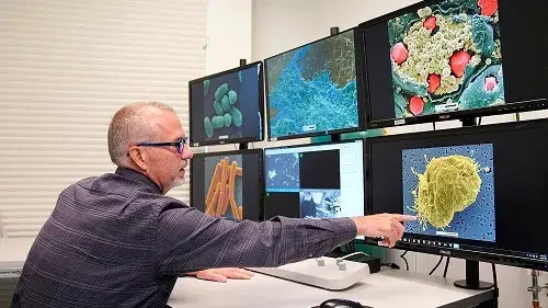
NBACC scientist using a scanning electron microscope points out a digital micrograph of Ebola virus coming out of a cell.
-
Image
NBACC scientist working on infectious samples in a self-contained Class III biosafety cabinet within a Biosafety Level 4 cabinet laboratory.
-
Image
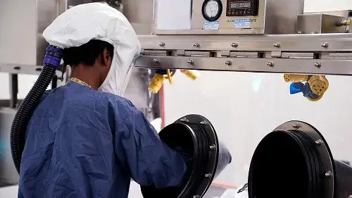
NBACC scientist working on infectious samples in a self-contained Class III biosafety cabinet within a Biosafety Level 4 cabinet laboratory.
-
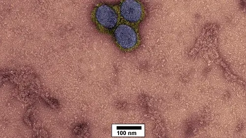
Image
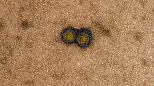
Negative stain transmission electron micrograph of SARS-CoV-2 (gold capsid, blue surface proteins). Magnification: 200,000X.
-
Image
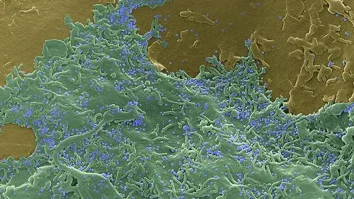
Scanning electron micrograph of the surface of a SARS-CoV-2 infected Vero cell (green), surrounded by less infected Vero cells (tan). SARS-CoV-2 can be seen budding from the surface and bound to the surface (blue). Magnification: 16,750X.
-
Image

Transmission electron micrograph of a section of a Vero cell (grey) with SARS-CoV-2 (blue capsid, yellow surface proteins) inside a vesicle (green). Magnification: 260,000X.
-
Image
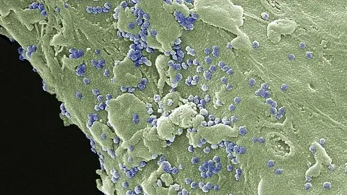
Scanning electron micrograph of the surface of a Vero cell (green) with SARS-CoV-2 on the cell surface (purple). Magnification: 40,000X.
-
Image
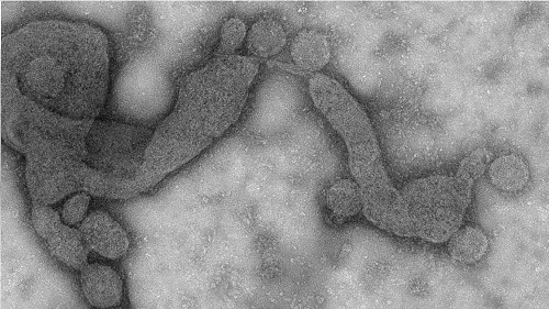
Negative stain transmission electron micrograph of SARS-CoV-2. Magnification: 150,000X.
-
Image
Negative stain transmission electron micrograph of SARS-CoV-2 (blue capsid, yellow surface proteins). Magnification: 200,000X.